

Images for illustrative purposes
Images for illustrative purposes
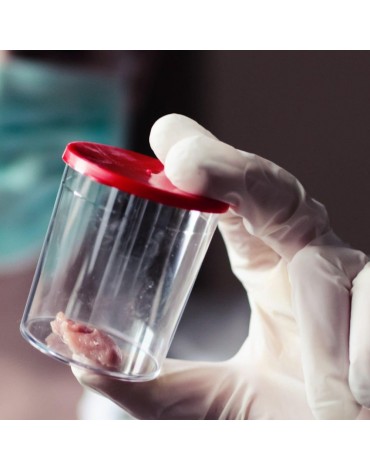
Pleural biopsy is a minimally invasive procedure, to remove a sample of the tissue that lines the lungs and inside the chest wall to detect disease or infection.
A local pain medicine (anesthetic) is injected. The doctor makes a small incision in the skin. The biopsy needle is inserted into abnormal tissue, tumor, or lung tissue. A small sample of tissue is removed with the needle.
Pleural biopsy is usually done to find the cause of a buildup of fluid around the lung (pleural effusion) or another abnormality of the pleural membrane. With pleural biopsy, tuberculosis, cancer, and other diseases can be diagnosed.